Get in touch
If you have any questions, just fill in the contact form or mail us directly at hello@gynaecam.com. We will get back to you shortly.
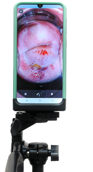
High resolution images of the cervix captured externally with a vivid color reproduction to spot anomalies.
Battery powered, bright embedded lighting within the device for a detailed image/video capture. Built in green filter.
Once fully charged, has a continuous working capacity of atleast 8 hours.
Easy to pack and transport. The design is built to last. Comes with a free waterproof travel bag.
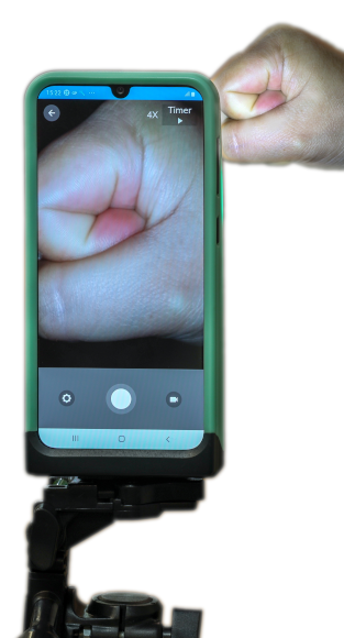
Enhanced ease of handling and stability during usage. Handheld and also available with a tripod.
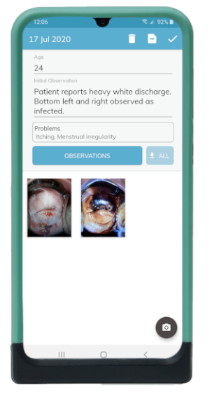
Store images on the device or the cloud, for remote diagnosis or future reference.
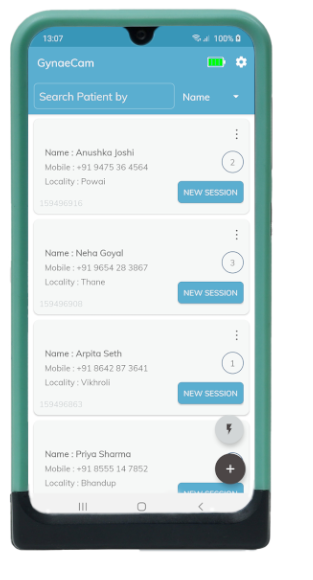
Access patient records anywhere, anytime. Tag important images and generate reports automatically. Quick access to previous records.


The primary inspiration behind GynaeCam’s development at IIT Bombay has been to develop an early detection tool for cervical cancer. Cervical cancer is the fourth most common cancer in women with approximately 570,000 cases being reported in 2018, out of which 311,000 died. India accounts for 97,000 of all cases reported worldwide, with 60,000 of those resulting as fatality.
Cervical cancer has a natural progression of 10-20 years, starting from the appearance of abnormal cells on the surface of the cervix, to full blown carcinoma cervix. This makes cervical cancer relatively easy to detect in its early stages, and further strengthens its case for early screening and treatment.
GynaeCam has been designed as a task shifting platform for cervical cancer screenings. GynaeCam follows IARC protocols for diagnosis. In its current form, our device is capable of assisting the diagnosis and documentation of several other gynaecological aliments.
If you have any questions, just fill in the contact form or mail us directly at hello@gynaecam.com. We will get back to you shortly.